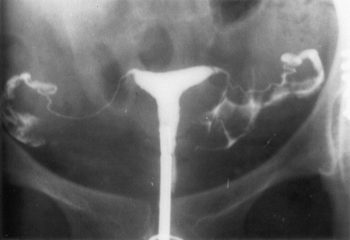
Hysterosalpingography (HSG)
Hysterosalpingography (HSG) refers to the method of X-ray diagnostics of the condition of the fallopian tubes and the internal cavity of the uterus, their patency and structure by introducing a contrast agent into the uterine cavity and tubes. This test is usually performed at the end of the menstrual cycle and before ovulation.
Endometrium biopsy Endometrium biopsy of the uterus is a procedure during which the sampling of the mucous membrane of the uterus - the endometrium is performed. An endometrium biopsy controls how the inner wall of the uterus reacts to hormones. This procedure not only provides information on the state of the endometrium, the presence of inflammation, and also determines whether progesterone is prepared for attachment of the embryo. Using a small catheter inserted through the cervix, tissue samples are taken for examination under a microscope. This test is usually done two to four days before the of menstruation.
Ultrasound This imaging tool takes images of internal organs using high frequency sound waves.. This procedure is performed in any period of the menstrual cycle, observe the development of follicles and ovulation. With ultrasound, the thickness of the cysts, fibroids and inner wall of the uterus are also checked.
Hysteroscopy For examination of the uterine cavity from the inside, a method such as hysteroscopy is used. Timely diagnosis allows you to determine the nature of uterine bleeding, the causes of infertility, the presence of diseases in uterus. Hysteroscopic examination is carried out using a special medical optical device - a hysteroscope. The heteroscope is a thin long rod with a backlight and a video camera.
Laparoscopy Laparoscopy is a minimal (minimally invasive) surgical intervention that allows you to diagnose certain diseases of the abdominal cavity, to explore the area of the uterus, fallopian tubes and ovaries. The advantage of laparoscopy is low traumatism, high efficiency, in a short recovery period and a minimum percentage of complications. During this procedure, because anesthesia is used, you will not feel any pain. The procedure can be performed at any time of the menstrual cycle.
Hyperechic focus in the heart Between the 18th and 30th weeks of pregnancy, ultrasound scanning can reveal a hyperechoic focus in the heart, popularly known as “Calcification of the heart”. It used to be assumed that an echogenic cardiac focus is associated with genetic diseases, such as Down syndrome
Hyperechoic focus is a term that speaks of the increased echogenicity (brightness) of a small area of the heart muscle on an ultrasound image. The detection of a hyperechoic focus in the heart is NOT a defect in the development of the heart, but simply reflects the nature of its ultrasound image. Hyperechoic focus occurs in the place of increased deposition of calcium salts on one of the heart muscles, which does not interfere with the normal operation of the fetal heart and does not require any treatment.


